Opened 2 months ago
Closed 2 months ago
#20346 closed defect (duplicate)
opening cryoEM SPA maps using "volume" view mode
| Reported by: | Owned by: | Tom Goddard | |
|---|---|---|---|
| Priority: | normal | Milestone: | |
| Component: | Volume Data | Version: | |
| Keywords: | Cc: | ||
| Blocked By: | Blocking: | ||
| Notify when closed: | Platform: | all | |
| Project: | ChimeraX |
Description
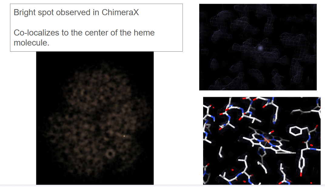
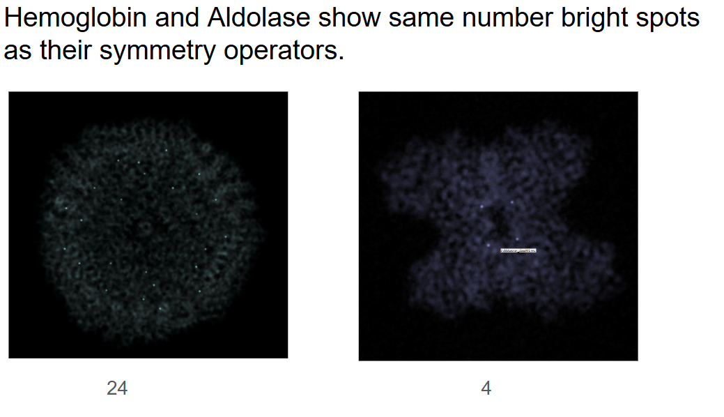
Hello wonderful people that build and maintain Chimera!


I have run into a bit of an odd observation which when compounded with some
basic biochemistry has left me scratching my head.   Essentially, when I
open one of our SPA volumes in chimerax and choos "volume" in the volume
viewer I am able to see a single bright spot in the volume.   If I process
our data with C2 symmetry we see two bright spots.

I had thought this was biologically relevant but I opened up a couple more
structures from our lab and other labs and see this is pretty common.
 Please see the attached images and you can see ApoFerritin has 24 bright
spots, and Aldolase has 4.

In the case of my sample - hemoglobin - the bright spot co-localizes with a
iron element in the structure.  I want to believe this has something to do
with the density/scattering of the Fe but am at a loss for why we would not
see 4 of them.   Hemoglobin has 4 heme centers and 4 Fe atoms.

Thank you in advance for any suggestions or support you can provide.

Thanks,
Remis


[image: image.png]


[image: image.png]


--

Jonathan Remis
*Scientific Engineer *
*Müller Lab*
*University of California, Berkeley*


Attachments (2)
Change History (4)
by , 2 months ago
comment:1 by , 2 months ago
| Component: | Unassigned → Volume Data |
|---|---|
| Owner: | set to |
| Platform: | → all |
| Project: | → ChimeraX |
| Status: | new → assigned |
comment:2 by , 2 months ago
| Resolution: | → duplicate |
|---|---|
| Status: | assigned → closed |
Forwarded to chimerax-users list for answering there.
Note:
See TracTickets
for help on using tickets.
Added by email2trac