How to Obtain Clear Views of Objects in Tomograms

Tom Goddard
January 23, 2009

Outline


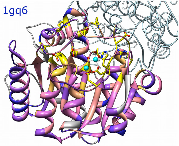
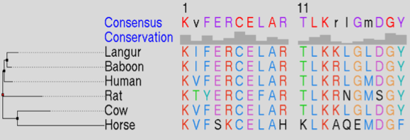
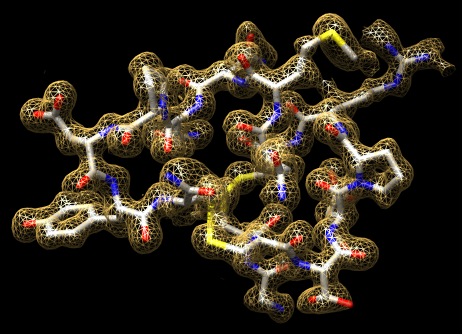
Atomic models and sequences

Morphing between conformations.

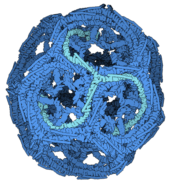
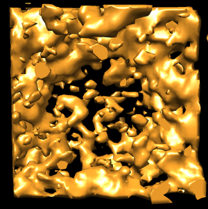
Molecular assemblies

Crystallography maps

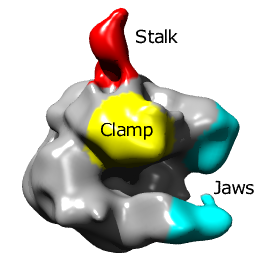
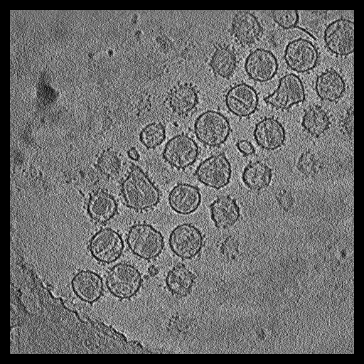
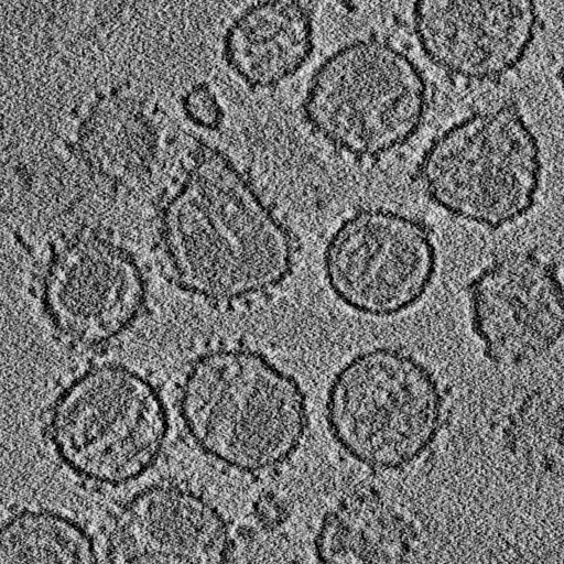
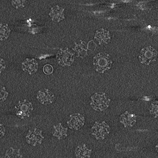
Single particle reconstructions
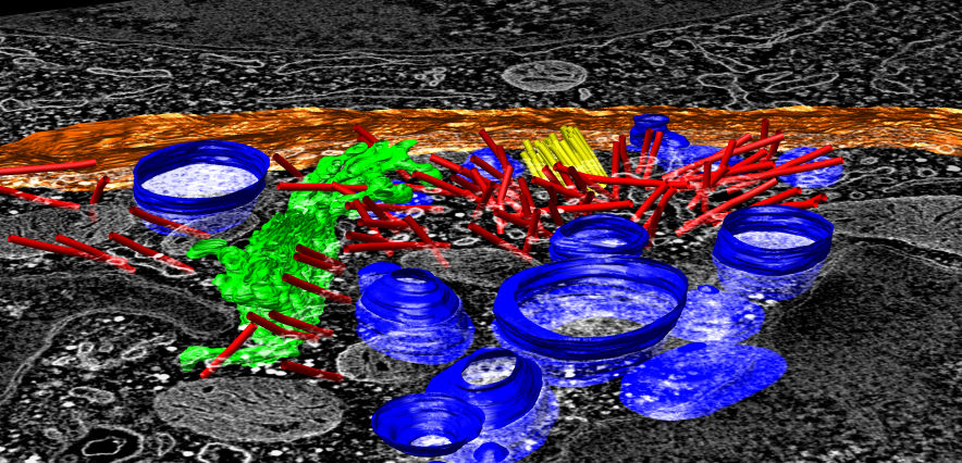
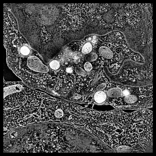
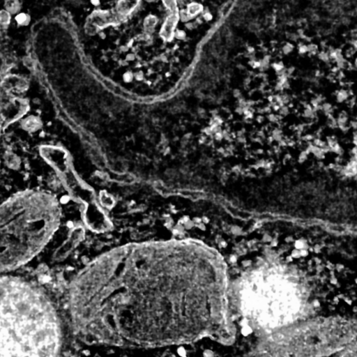
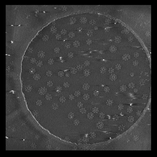
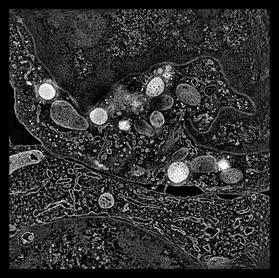
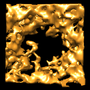
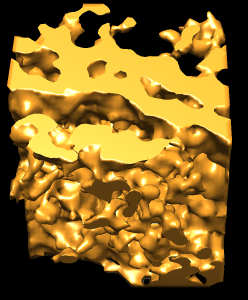
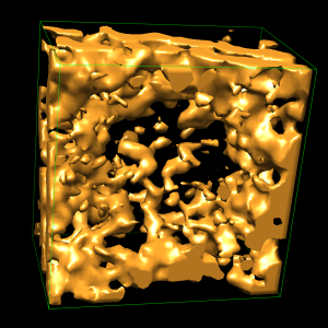
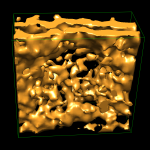
EM tomography

Nuclear Pores

SIV Virus Spikes

Clathrin Coated Vesicles

Common Goal

Common Problems

Common Features

Demonstration

  • Point out human T-cell outline.
  • Point out nuclear envelope.
  • Flip through planes and mark 3 pores. 250 A diameter, color blue.
  • Use volume dialog atom box to show box around center marker. 500 A pad.
  • Surface display style.
  • Color orange to see white specular highlights better.
  • Hide dust. Move slider to show effect.
  • For unstained samples that have more dust this is especially useful.
  • Lower threshold obscures view.
  • Adjust box tighter (and wider) so less density blocks view.
  • Rotate box to align with pore, allows tighter cropping.
  • Rotating makes a new map interpolated from original map on rotated grid.
  • Extract circular region of pore to examine 8-fold symmetry.
  • Center marker, color zone, split map. 450 A radius.
  • Gaussian filter pore.
  • Use interactive update and adjust Gaussian width with slider. 45 A good.
  • Find symmetry axis of pore.
  • Close unneeded maps, duplicate gaussian map, rotate by hand ~180, fit map in map, "measure rotation #2 #3".

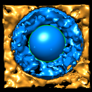
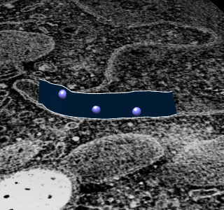
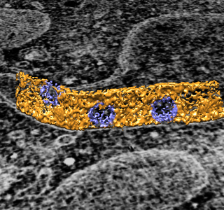
To extract density new many membrane embedded objects like 100 virus spikes rotating a box around each is time consuming. Better to extract density near membrane surface for the entire membrane.

  • Trace nuclear envelope surface.
  • Set mouse mode, new marker set, trace top/bottom plane curves, surface.
  • "mask #0 sel slab 700", show as surface

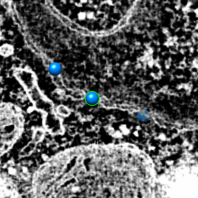
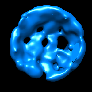
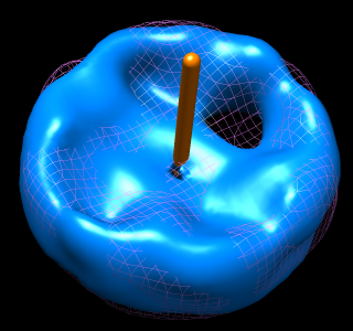
To show pores in their environment make a fly-through animation.

movie

Chimera Setup and Support