Chimera Tutorial:
Visualizing Electron Tomography

Tom Goddard
July 12, 2012

This tutorial covers basic techniques for viewing, filtering, and segmenting noisy electron tomography maps using Chimera 1.6.

We will look at a map of HIV virus particles from the Fuller lab, EM Databank 1155.

Displaying XY Planes Inverting Intensity Values
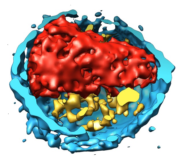
  1. Open map emd_1155.map.
  2. Volume dialog, Features / Planes, press One button.
  3. Drag markers on histogram brightness yellow curve.
  4. Move Plane slider to flip through planes.
  5. Change plane axis to z and flip through planes.
  6. Show all planes.
  1. Note small map values are high density. Want large map values for high density.
  2. Volume dialog, Tools / Volume Filter, type Scale, scale -1, options turn off displayed subregion only, press Filter.
  3. Hide original map by clicking "eye" icon above histogram.
  4. Switch from surface style to solid, One plane.
Hide Dust Gaussian Filtering
  1. Hide small surface blobs to reduce noise.
  2. Volume style surface, All planes.
  3. Volume dialog, Tools / Hide Dust, Surface emd_1155.map scaled, press Hide.
  4. Drag vertical bar on volume dialog histogram to lower contour level.
  5. Move size slider in hide dust dialog.
  6. Press Unhide.
  1. Smooth map to reveal large-scale features.
  2. Volume dialog, Tools / Volume Filter, type Gaussian, width 30 Angstroms, value type float32, select scaled map in volume dialog, press Filter.
  3. Planes One, depth 10, move plane slider.
Extract One Virus Mask Virus Membrane
  1. Volume dialog, Features / Subregion Selection, enable "Select subregions", drag box around middle virus particle, press Crop.
  2. Click on green outline box face (not edge) and drag. Press Crop.
  3. Uncheck "Select subregions" to move map using mouse.
  4. Can save map to new file with volume dialog, File / Save Map As....
  1. Show command line using Favorites / Command Line
  2. Enter command "shape sphere radius 550".
  3. Click off "Active" sphere model #3 below command-line to stop mouse from moving sphere.
  4. Move map to be centered on sphere. Ctrl-middle-mouse moves in z direction.
  5. Mesh display style can help see sphere inside map.
  6. Create map for inside of sphere, command "mask #2 #3"
  7. Hide sphere, Favorites / Model Panel, uncheck shown button for sphere.
  8. Create map ouside sphere, command "mask #2 #3 invert true".
  9. Color inside map red, command "volume #4 color red".
Clip Virus Membrane
  1. Cut virus membrane in half to see inside.
  2. Tools / Depiction / Per-Model Clipping, Model #5, check "Enable clipping".
  3. Check "Adjust clipping with mouse", move clip plane by dragging with middle mouse button in graphics window.