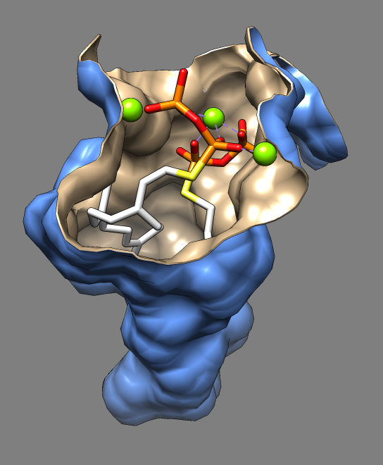
Binding pocket of dehydrosqualene synthase from Staphylococcus aureus with an inhibitor farnesyl thiopyrophosphate that reduces virulence in hospital acquired infections. This image illustrates a few tricks aimed at making clearer depictions of binding site surfaces. The example comes from the Chimera structure comparison tutorial. Two superimposed surfaces enable different colors on the inside and outside. The surface has thickness (0.1 Å) as seen at the surface edge. The surface edge is smoothed by cutting triangles of the original molecular surface in half. Detached patches of surface that were floating near the ligand were hidden. These effects were achieved by creative use of the "measure contactArea" command to show the contact between the ligand surface and the receptor surface, in combination with the "sop hideDust" command to remove the stray surface patches. See the example command script for details.
Chimera session file using Chimera 1.6 (September 19, 2011 build).
© 2009 The Regents of the University of California; all rights reserved.